

Contents
Scroll to:
 E. O. Ionova,
I. B. Tsorin,
M. B. Vititnova,
V. N. Stolyaruk,
I. M. Roshchevskaya,
S. A. Kryzhanovskii
E. O. Ionova,
I. B. Tsorin,
M. B. Vititnova,
V. N. Stolyaruk,
I. M. Roshchevskaya,
S. A. Kryzhanovskii https://doi.org/10.37489/10.37489/2587-7836-2026-1-73-79
EDN: UIVRZJ
Scroll to:
Objective. To study the features of changes in the geometry of the left ventricle of the heart and its inotropic function in the acute phase of myocardial infarction (MI) in rats with established alcoholic cardiomyopathy (ACMP).
Materials and methods. The experiments were carried out on a translational model of ACMP in rats that we developed, according to which this pathology is formed after 24 weeks of forced intake of 10 % ethanol solution. MI was reproduced by one-stage ligation of the descending coronary artery 1–2 mm below the place of its exit from under the left atrial appendage.
Results. It was shown that in rats with ACMP, already in the first minutes after the reproduction of acute myocardial infarction, the heart loses its physiological shape and becomes spherical. The change in the geometry of the heart occurs against the background of a significant (p = 0.001) decrease in the inotropic function of the left ventricle by ≈ 30 %, while in control animals the decrease is ≈ 15 %.
Conclusion. In animals with formed AKMP, myocardial infarction is more severe than in intact animals, in particular, they have a fairly high risk of developing an aneurysm of the left ventricle of the heart and, consequently, the risk of developing severe, progressive heart failure with a high probability of sudden cardiac death.
Ionova E.O., Tsorin I.B., Vititnova M.B., Stolyaruk V.N., Roshchevskaya I.M., Kryzhanovskii S.A. Study of the state of contractile function of the heart and the dynamics of changes in its geometry in the acute phase of anterior transmural myocardial infarction in rats with alcoholic cardiomyopathy. Pharmacokinetics and Pharmacodynamics. 2026;(1):73-79. (In Russ.) https://doi.org/10.37489/10.37489/2587-7836-2026-1-73-79. EDN: UIVRZJ
According to the WHO report “Global Burden of Disease” for 2019, alcohol abuse is a significant risk factor in terms of disability-adjusted life years (DALYs), accounting for 3.7% of DALYs in all age groups, and this indicator increased by 37% from 1990 to 2019 [1]. It has also been shown that alcohol abuse is the main risk factor for disability in people aged 25 to 49 years and represents the first preventable cause of disease in this age group [1]. The most common somatic complication caused by chronic alcohol abuse is alcoholic cardiomyopathy (ACMP) — an acquired form of dilated cardiomyopathy, which is the leading cause of death (50–60% of cases) in patients with chronic alcoholism [2]. ACMP accounts for approximately 35% of all non-ischemic cardiomyopathies [3]. In chronic alcoholics with established ACMP, mortality during continued alcohol consumption is significantly higher than in patients with idiopathic dilated cardiomyopathy [4]. It should be emphasized that sudden cardiac death (SCD) accounts for 30–40% of ACMP mortality [5, 6]. Among all causes of sudden cardiac death, ACMP ranks second, second only to acute coronary syndrome (ACS) [7, 8]. Moreover, ACMP is the most common cause of non-ischemic SCD in patients aged 40 to 59 years [6]. Data on the contribution of ischemic heart disease, in particular ACS, to the development of SCD in patients with established ACMP have not yet been presented in the literature; however, there are reports that among those who died from ACMP, 8.4% were diagnosed with coronary stenosis with vessel lumen reduction of at least 50% [9]. It cannot be ruled out that one of the possible causes of SCD in patients with established ACMP may be ACS, namely its acute phase.
The aim of this study was to investigate the features of changes in left ventricular geometry and its inotropic function in the acute phase of myocardial infarction in rats with established ACMP.
Animals. Experiments were performed on white outbred male rats with an initial weight of 180–200 g, obtained from the “Scientific Center of Biomedical Technologies of the Federal Medical and Biological Agency”, branch “Stolbovaya”, having a veterinary certificate of quality and health, and having undergone a 15-day quarantine in the vivarium of the Federal Research Center for Innovator and Emerging Biomedical and Pharmaceutical Technologies. Animals were kept in individual standard plastic cages, with pelleted feed provided ad libitum under a regulated 12/12 light cycle (light off at 08:00 am). Animal housing conditions complied with GOST 33215–2014 “Guidelines for housing and care of laboratory animals. Rules for equipment of premises and organization of procedures” (Reissue) and GOST 33216–2014 “Guidelines for housing and care of laboratory animals. Rules for housing and care of laboratory rodents and rabbits” (Reissue). All work with laboratory animals was performed in accordance with generally accepted norms for animal handling, based on standard operating procedures adopted at the Federal Research Center for Innovator and Emerging Biomedical and Pharmaceutical Technologies, international rules (European Communities Council Directive of November 24, 1986 (86/609/EEC)), and the “Rules for Working with Animals” approved by the Bioethics Committee of the V.V. Zakusov Research Institute of Pharmacology. Animals received standard pelleted feed PK-120-1 (Laboratorsnab LLC, Russian Federation).
Study design. Animals were randomized into 2 groups: 1st (n = 8) — control (myocardial infarction in intact rats), 2nd (n = 10) — ACMP (myocardial infarction in rats with ACMP). Before the experiment, animals were anesthetized (urethane 1300 mg/kg, i.p.) and placed in the supine position on a heated table for small laboratory animals “Surgi Suite” (Kent Scientific Corporation, USA). Then the animals were placed on artificial ventilation using a ventilator “Model 7025” (Ugo Basile, Italy).
Method for reproducing ACMP. Experiments were performed on our previously developed translational model of ACMP in rats [10]. To model ACMP, animals were subjected to forced alcoholization for 6 months by providing a 10% ethanol solution as the only source of fluid. The average daily alcohol consumption in terms of pure ethanol ranged from 5.0 to 6.5 g/kg. It has been shown that after 24 weeks of forced alcoholization, animals develop alcoholic cardiomyopathy, reproducing the main clinical and diagnostic signs of this disease.
Method for reproducing acute myocardial infarction (AMI). AMI was reproduced by one-stage ligation of the descending coronary artery 1–2 mm below its exit from under the left atrial appendage.
Echocardiographic examination. Measurements were performed under open-chest conditions in one-dimensional M-mode and two-dimensional B-mode with the echocardiograph transducer positioned in the parasternal long-axis view of the heart. In M-mode, left ventricular end-systolic (ESD) and end-diastolic (EDD) dimensions were assessed. Then, using the Teichholz method, the ejection fraction (EF), an indicator of cardiac contractile function, was calculated. In addition, such parameters as anterior wall thickness in systole (AWTs) and diastole (AWTd), posterior wall thickness in systole (PWTs) and diastole (PWTd), anterior wall systolic thickening (AWST), and cardiac index were evaluated. Echocardiographic parameters were assessed over at least five consecutive cardiac cycles. All measurements were performed in accordance with the Recommendations of the American Society of Echocardiography and the European Association of Echocardiography [11]. A digital ultrasound echocardiograph “DC-60” (Mindray, China) with an electronic phased array transducer P10-4E (5.0/11.0 MHz) was used.
Statistical analysis. Normality of distribution was tested using the Shapiro–Wilk test, homogeneity of variances using Levene’s test. Since the sample distributions were close to normal and variances were homogeneous, repeated measures analysis of variance followed by Duncan’s multiple comparison test was used to determine statistical significance. Results are presented as arithmetic means and their standard errors. Changes were considered statistically significant at a type I error probability of p ≤ 0.05.
As a certified model of ACMP in rats, we used our previously developed translational model of this pathology [10], which has gained recognition and is cited in the Western literature [12]. According to the results of echocardiographic studies, in rats that had consumed 10% ethanol solution forcibly for 24 weeks, left ventricular (LV) ESD and EDD increased compared to control animals by 36.5% (p = 0.008) and 15.2% (p = 0.017), respectively, while EF, characterizing the inotropic status of the left ventricle, significantly (p = 0.001) decreased from 89.0±1.8 to 78.1±1.5 (Table 1). The recorded changes indicate the formation in the main group of rats of echocardiographic signs of alcohol-induced dilated ACMP (LV dilatation and decreased contractility), which is almost completely consistent with those known for humans with this pathology [13]. It is known that in a healthy adult at rest, EF averages 50–60% [14].
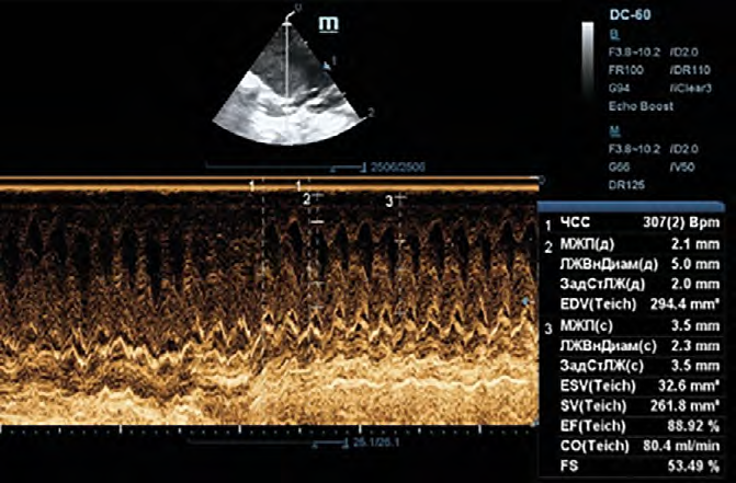
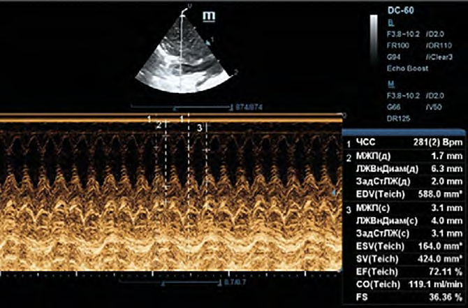
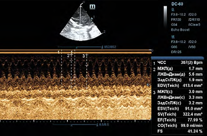
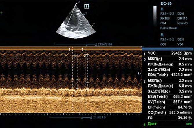
When planning this series of experiments, we expected to show what seemed obvious to us — that acute myocardial infarction in rats with ACMP is significantly more severe from the first minutes of its onset than in control animals without ACMP. However, the obtained results, at first glance, did not correspond to this concept. For example, in control rats by the 15th minute of ischemia, ESD increased by 63% (from 2.46±0.17 mm to 4.03±0.30 mm), whereas in rats with ACMP it increased by only 45% (from 3.41±0.16 to 4.94±0.27). The same applies to EF — in control rats by the 15th minute of ischemia, it decreased by 19% (from 89.0±1.8% to 72.2±2.5%), and in rats with ACMP by 15% (from 78.1±1.5% to 66.6±2.1%). A similar picture was observed at the 30th minute of ischemia (Fig. 1, Table 1).
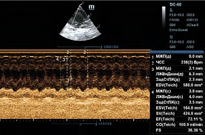
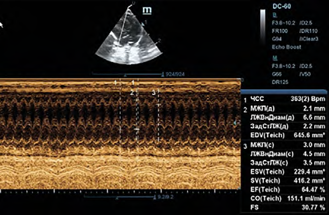
However, subsequent analysis of the results disproved the initial conclusion and led us to think about the possible life‑compatible magnitudes of pathological changes in heart geometry. In our case, myocardial infarction in rats with ACMP was reproduced against the background of established ACMP, i.e., against the background of alcohol‑induced chronic heart failure. In these animals, before the induction of myocardial infarction, ESD was already increased by ≈40%, i.e., the actual increase in ESD by the 15th minute of ischemia was close to 90% (Table 1). A similar calculation shows that in rats with ACMP the actual increase in EDD is 50%, whereas in intact rats it is more than two times less (19%). Similarly, all recorded parameters reflecting left ventricular geometry actually change (Table 1), suggesting that in animals with ACMP, already in the first minutes after the induction of acute myocardial infarction, the heart loses its physiological shape and becomes spherical, which is hemodynamically extremely unfavorable.
These geometric changes occur against the background of an actual decrease in LV EF (inotropic function) — by 25% at the 15th minute of ischemia and by 30% at the 30th minute. Such a significant decrease in EF is apparently associated with the loss of contractile ability of the anterior wall of the left ventricle: whereas in control rats during systole the anterior wall contracts almost as well as before coronary ligation — its thickening by the 30th minute of ischemia decreases by only 9%, in rats with ACMP this decrease is 58% — from 68.4±4.6% to 29.3±4.5% (Table 1). Such a drop in anterior wall contractility during systole is prognostically unfavorable, as it indicates a high risk of both left ventricular aneurysm formation and cardiac rupture. It is known that in a healthy adult at rest, EF averages 50–55–60% [14]. If we extrapolate the magnitude of EF reduction to humans, then by the 30th minute of ischemia, EF would be ≈35–40%, which, according to the 2020 Clinical Practice Guidelines of the Russian Society of Cardiology “Chronic Heart Failure”, is classified as heart failure with reduced ejection fraction (≤40%), and is also prognostically unfavorable [15]. Low EF, as well as the degree of LV dilatation, are considered possible predictors of SCD, and the larger the LV diameter and/or the lower the EF, the potentially higher the risk of SCD [16, 17]. For example, according to the Maastricht registry (more than 180,000 participants), circulatory arrest due to SCD was recorded in patients with EF ≥50% in 1.4% of cases, with EF 31–40% in 5.1% of cases, and with EF ≤30% in 7.5% of cases [18].
 |  |  |
| Background control | Control 15 min | Control 50 min |
 |  |  |
| Background ACMP | ACMP 15 min | ACMP 50 min |
Fig. 1. Original echocardiograms of rat No. 5 from the control group (top row) and rat No. 12 with ACMP (bottom row). From left to right: baseline before coronary artery ligation and 15 and 30 minutes after ligation
Table 1. Changes in contractile function and geometry of the heart occurring in the acute phase of anterior transmural myocardial infarction in anesthetized rats (urethane 1300 mg/kg i.p.) with established alcoholic cardiomyopathy
| Parameter | Group | n | Baseline before coronary ligation | 15 min coronary occlusion | 30 min coronary occlusion |
|---|---|---|---|---|---|
| LV end-systolic dimension, mm | Control | 8 | 2.46±0.17 | 4.03±0.30 p < 0.001 | 4.00±0.25 p < 0.001 |
| ACMP | 10 | 3.41±0.16 p1 = 0.008 | 4.94±0.27 p < 0.001; p1 = 0.026 | 4.74±0.22 p <0.001; p1 = 0.083 | |
| PWT systole, mm | Control | 8 | 3.63±0.07 | 3.33±0.09 p < 0.001 | 3.34±0.08 p < 0.001 |
| ACMP | 10 | 3.46±0.10 p1 = 0.19 | 3.39±0.08 p = 0.30; p1 = 0.62 | 3.39±0.09 p = 0.27; p1 = 0.69 | |
| AWT systole, mm | Control | 8 | 3.66±0.11 | 3.08±0.18 p < 0.001 | 3.08±0.15 p < 0.001 |
| ACMP | 10 | 3.53±0.10 p1 = 0.52 | 2.77±0.16 p < 0.001; p1 = 0.16 | 2.66±0.14 p < 0.001; p1 = 0.06 | |
| LV end-systolic volume, mm³ | Control | 8 | 42.5±12.6 | 181.6±43.3 p < 0.001 | 177.9±28.9 p < 0.001 |
| ACMP | 10 | 107.0±11.2 p1 = 0.001 | 308.8±38.7 p < 0.001; p1 = 0.005 | 265.6±25.9 p < 0.001; p1 = 0.051 | |
| LV end-diastolic dimension, mm | Control | 8 | 5.33±0.21 | 6.34±0.36 p < 0.001 | 6.58±0.34 p < 0.001 |
| ACMP | 10 | 6.09±0.19 p1 = 0.017 | 7.44±0.33 p < 0.001; p1 = 0.021 | 7.15±0.30 p < 0.001; p1 = 0.18 | |
| PWT diastole, mm | Control | 8 | 2.36±0.06 | 2.30±0.07 p = 0.12 | 2.23±0.06 p = 0.002 |
| ACMP | 10 | 2.23±0.06 p1 = 0.18 | 2.19±0.05 p = 0.34; p1 = 0.28 | 2.13±0.05 p = 0.023; p1 = 0.34 | |
| AWT diastole, mm | Control | 8 | 2.35±0.08 | 2.01±0.12 p = 0.011 | 2.04±0.11 p = 0.017 |
| ACMP | 10 | 2.10±0.08 p1 = 0.086 | 2.05±0.07 p = 0.68; p1 = 0.81 | 2.06±0.07 p = 0.73; p1 = 0.88 | |
| AWST, % | Control | 8 | 57.2±5.1 | 54.3±5.3 p = 0.53 | 52.5±5.0 p = 0.34 |
| ACMP | 10 | 68.4±4.6 p1 = 0.11 | 34.5±4.7 p < 0.001; p1 = 0.010 | 29.3±4.5 p < 0.001; p1 = 0.003 | |
| LV end-diastolic volume, mm³ | Control | 8 | 374.0±48.1 | 625.3±112.0 p = 0.007 | 695.9±89.2 p = 0.001 |
| ACMP | 10 | 534.2±43.0 p1 = 0.024 | 948.9±100.2 p < 0.001; p1 = 0.015 | 837.1±79.8 p = 0.002; p1 = 0.24 | |
| LV fractional shortening, % | Control | 8 | 53.9±2.0 | 37.1±2.6 p < 0.001 | 39.8±1.4 p < 0.001 |
| ACMP | 10 | 41.8±1.6 p1 < 0.001 | 32.6±2.0 p < 0.001; p1= 0.085 | 33.5±1.4 p < 0.001; p1 = 0.018 | |
| LV ejection fraction, % | Control | 8 | 89.0±1.8 | 72.2±2.5 p < 0.001 | 75.7±1.6 p < 0.001 |
| ACMP | 10 | 78.1±1.5 p1 < 0.001 | 66.6±2.1 p < 0.001; p1 = 0.050 | 62.0±1.7 p < 0.001; p1 = 0.011 | |
| Heart rate, beats/min | Control | 8 | 234±16 | 293±16 p < 0.001 | 298±21 p < 0.001 |
| ACMP | 10 | 238±15 p1 = 0.87 | 303±15 p < 0.001; p1 = 0.071 | 304 p < 0.001; p1 = 0.082 | |
| Cardiac index, mL/min/kg | Control | 8 | 135.5±19.0 | 220.1±43.9 p = 0.019 | 263.0±34.4 p < 0.001 |
| ACMP | 10 | 186.7±17.0 p1 = 0.061 | 359.4±39.2 p < 0.001; p1 = 0.007 | 317.0±30.7 p < 0.001; p1= 0.25 |
Notes: 1) Arithmetic means and their standard errors are shown; 2) p — compared to baseline; 3) p1 — compared to intact rats; 4) Abbreviations: PWT – posterior wall thickness, AWT – anterior wall thickness, AWST – anterior wall systolic thickening.
Thus, the data obtained in this series of experiments allow us to state with a certain degree of confidence that in animals with established ACMP, myocardial infarction is more severe than in intact animals; in particular, they have a fairly high risk of developing left ventricular aneurysm and, consequently, the risk of developing severe, progressive heart failure with a high probability of sudden cardiac death.
1. GBD 2019 Risk Factors Collaborators. Global burden of 87 risk factors in 204 countries and territories, 1990-2019: a systematic analysis for the Global Burden of Disease Study 2019. Lancet. 2020 Oct 17;396(10258):12231249. doi: 10.1016/S0140-6736(20)30752-2.
2. Bohan NA, Mandel AI, Maximenko NN, Mihaleva LD. Lethal outcomes in alcoholism. Narcology. 2007;12:37-40. (In Russ.).
3. Figueredo VM, Patel A. Detrimental Effects of Alcohol on the Heart: Hypertension and Cardiomyopathy. Rev Cardiovasc Med. 2023 Oct 17;24(10):292. doi: 10.31083/j.rcm2410292.
4. Fauchier L, Babuty D, Poret P, et al. Comparison of long-term outcome of alcoholic and idiopathic dilated cardiomyopathy. Eur Heart J. 2000 Feb;21(4):306-14. doi: 10.1053/euhj.1999.1761.
5. Corović N, Duraković Z, Misigoj-Duraković M. Dispersion of the corrected QT and JT interval in the electrocardiogram of alcoholic patients. Alcohol Clin Exp Res. 2006 Jan;30(1):150-4. doi: 10.1111/j.1530-0277.2006.00018.x.
6. Guzzo-Merello G, Segovia J, Dominguez F, et al. Natural history and prognostic factors in alcoholic cardiomyopathy. JACC Heart Fail. 2015 Jan;3(1):78-86. doi: 10.1016/j.jchf.2014.07.014. Epub 2014 Nov 11. PMID: 25458176.
7. Kovalev AV, Morozov IuE, Samokhodskaia OV, Bereznikov AV. Alcohol-associated mortality in Russia (based on the materials for the period from 2011 till 2016). Forensic Medical Expertise. 2017;60(6):4-8. (In Russ.). doi: 10.17116/sudmed20176064-8.
8. Hookana E, Junttila MJ, Puurunen VP, et al. Causes of nonischemic sudden cardiac death in the current era. Heart Rhythm. 2011 Oct;8(10):1570-5. doi: 10.1016/j.hrthm.2011.06.031.
9. Hietanen S, Herajärvi J, Junttila J, et al. Characteristics of subjects with alcoholic cardiomyopathy and sudden cardiac death. Heart. 2020 May;106(9):686-690. doi: 10.1136/heartjnl-2019-315534.
10. Kryzhanovskii SA, Kolik LG, Tsorin IB, et al. Alcoholic cardiomyopathy: Translation model. Bull Exp Biol Med. 2017;163(5):627-631. (In Russ.). doi: 10.1007/s10517-017-3865-0.
11. Lang RM, Bierig M, Devereux RB, et al; Chamber Quantification Writing Group; American Society of Echocardiography's Guidelines and Standards Committee; European Association of Echocardiography. Recommendations for chamber quantification: a report from the American Society of Echocardiography's Guidelines and Standards Committee and the Chamber Quantification Writing Group, developed in conjunction with the European Association of Echocardiography, a branch of the European Society of Cardiology. J Am Soc Echocardiogr. 2005 Dec;18(12):1440-63. doi: 10.1016/j.echo.2005.
12. Argo A, Pitingaro W, Puntarello M, et al. A Comprehensive Review on Alcohol Abuse Disorder Fatality, from Alcohol Binges to Alcoholic Cardiomyopathy. Diagnostics (Basel). 2024 Jun 5;14(11):1189. doi: 10.3390/diagnostics14111189.
13. Pinto YM, Elliott PM, Arbustini E, et al. Proposal for a revised definition of dilated cardiomyopathy, hypokinetic non-dilated cardiomyopathy, and its implications for clinical practice: a position statement of the ESC working group on myocardial and pericardial diseases. Eur Heart J. 2016 Jun 14;37(23):1850-8. doi: 10.1093/eurheartj/ehv727.
14. Lang RM, Badano LP, Mor-Avi V, et al. Recommendations for cardiac chamber quantification by echocardiography in adults: an update from the American Society of Echocardiography and the European Association of Cardiovascular Imaging. J Am Soc Echocardiogr. 2015 Jan;28(1):1-39.e14. doi: 10.1016/j.echo.2014.10.003.
15. 2020 Clinical practice guidelines for Chronic heart failure. Russian Journal of Cardiology. 2020;25(11):4083. (In Russ.). doi: 10.15829/1560-4071-2020-4083.
16. Buxton AE, Lee KL, Hafley GE, et al; MUSTT Investigators. Relation of ejection fraction and inducible ventricular tachycardia to mode of death in patients with coronary artery disease: an analysis of patients enrolled in the multicenter unsustained tachycardia trial. Circulation. 2002 Nov 5; 106(19):2466-72. doi: 10.1161/01.cir.0000037224.15873.83.
17. Narayanan K, Reinier K, Teodorescu C, et al. Left ventricular diameter and risk stratification for sudden cardiac death. J Am Heart Assoc. 2014 Sep 16;3(5):e001193. doi: 10.1161/JAHA.114.001193.
18. Santangeli P, Dello Russo A, Casella M, et al. Left ventricular ejection fraction for the risk stratification of sudden cardiac death: friend or foe? Intern Med J. 2011 Jan;41(1a):55-60. doi: 10.1111/j.1445-5994.2010.02371.x.1.
Ekaterina O. Ionova — PhD, Cand. Sci. (Med.), Leading Researcher of Laboratory of Circulation Pharmacology.
Moscow
Iosif B. Tsorin — PhD, Dr. Sci. (Biology), Leading Researcher of Laboratory of Circulation Pharmacology.
Moscow
Marina B. Vititnova — PhD, Cand. Sci. (Biology), Leading Researcher of Laboratory of Circulation Pharmacology.
Moscow
Valeriy N. Stolyaruk — PhD, Cand. Sci. (Med.), Leading Researcher of Laboratory of Circulation Pharmacology.
Moscow
Irina M. Roshchevskaya — PhD, Dr. Sci. (Biology), Professor, Corresponding Member RAS, Chief Researcher of Laboratory of Circulation Pharmacology.
Moscow
Sergey A. Kryzhanovskii — PhD, Dr. Sci. (Med.), Head of Laboratory of Circulation Pharmacology.
Moscow
Ionova E.O., Tsorin I.B., Vititnova M.B., Stolyaruk V.N., Roshchevskaya I.M., Kryzhanovskii S.A. Study of the state of contractile function of the heart and the dynamics of changes in its geometry in the acute phase of anterior transmural myocardial infarction in rats with alcoholic cardiomyopathy. Pharmacokinetics and Pharmacodynamics. 2026;(1):73-79. (In Russ.) https://doi.org/10.37489/10.37489/2587-7836-2026-1-73-79. EDN: UIVRZJ
НАШИ КНИГИ




Другие журналы
"Издательства ОКИ"


ПАРТНЕРЫ

 Email this article (Login required)
Email the author (Login required)
Email this article (Login required)
Email the author (Login required) Editorial address:
121357, Moscow, st. Vatutina, 4, building 2, apt. 22
General Director of OKI Publishing House LLC
Afanasyeva Elena
Tel. + 7 (916) 986-04-65; Email: eva88@list.ru
Processing of personal data


